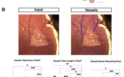
Traditional pathology utilizes stained blood smears viewed through microscopy, a technique still prevalent despite technological advancements. Typically, manual human assessment accompanies automated methods, maintaining simplicity but lacking comprehensive cell...
IKOSA AI
AI-Supported Quality Assurance in Stem Cell Bioprocessing: An Interview with Dr. Klaus Graumann
Welcome everybody to our interview. The topic will be “AI-supported quality assurance in stem cell bioprocessing”. Elisa and I are very excited to be speaking with Klaus Graumann today about the technological innovations and ongoing initiatives within his...
ROI, Annotation, Labels: Mastering Image Annotation in Pathology
Microscopy image annotation and pathology annotation, in particular, are tasks that require a lot of attention to detail. To be able to train Deep Learning Computer Vision Applications, which perform advanced tasks such as detection, segmentation, and classification...
How to Successfully Conduct Morphological Cell Profiling With The Cell Painting Assay
The Cell Painting Assay provides researchers with a powerful tool for morphological cell profiling, enabling investigations into cellular behavior and the effect of therapeutic compounds on cellular structures. In this article, we explore the principles and...
From Microscopic Patterns to Diagnostic Insights: Exploring the New Frontier of Blood and Inflammation with Prof. Johannes Schmid
Join us, as we embark on a captivating journey delving into the groundbreaking work and ongoing projects of Prof. Johannes Schmid from the Medical University of Vienna. In this interview, we shine a spotlight on the remarkable realm of "Multifactorial Fluorescence...
Toxicological Analysis by Assessment of Vascularization and Cell Viability Using the Chicken’s Chorioallantoic Membrane (CAM Assay)
This paper presents a promising alternative in animal experimentation - the CAM assay using hen's eggs. The CAM provides a suitable environment for xenograft implantation, enabling the growth of human tumors. Evaluating therapeutic agents for efficacy and toxicity...
Simplifying N:C Ratio Analysis in Cytology: An Easy Approach Using Image Analysis Software
In recent times histological assessment is considered the gold standard for assessing cell and tissue malignancy or the absence thereof. However, it is still a very time-consuming method, the results of which may strongly depend on differing interpretations from...
Spatial Biology: Introducing Spatial Metrics to Bioimage Analysis
Spatial biology is a growing field in modern life science with a significant future potential. We dive deep into this exciting subject to keep you updated with the latest developments in the field. Find out about state-of-the art spatial biology...
Pathologist Advice on Prostate Cancer Microscopy Analysis Techniques
Prostate microscopy is one of the areas where the use of pathology image analysis is on the rise and about to become standard diagnostic practice. In this article, you will learn about the recent developments in automated prostate tissue analysis. Find out how...
Unlocking the Full Potential of Multiplex IHC Analysis
Shifting from single marker analysis to more complex multiplex IHC analysis methods is an important step towards improving the outcomes of immunohistochemistry studies. Yet, different multiplex immunohistochemistry (mIHC) techniques generate tons of image data for...
Join our newsletter
Be the first to receive free tips about image analysis, newest articles and publications!