In this article, we introduce five angiogenesis quantification software solutions that will help you harness the potential of AI-driven image analysis in vasculogenesis research. Find out how state-of-the-art computer vision technology will assist you in the study of blood vessel growth processes. Using these advanced software applications you can easily evaluate vascular development and angiogenesis inhibition based on different quantitative parameters.
- Understanding the angiogenic blood vessel growth process
- The role of vascular endothelial growth factors (VEGF) in angiogenic processes
- The role of angiogenic markers in angiogenesis quantification
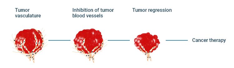
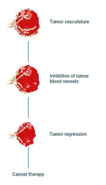
- Angiogenesis inhibition in cancer treatment
- How angiogenesis research benefits from automated image analysis software
- Unveiling the IKOSA portfolio of specialized solutions for the analysis of angiogenesis images
Understanding the angiogenic blood vessel growth process
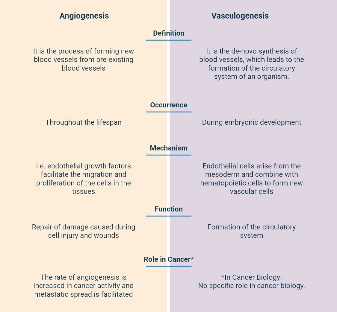
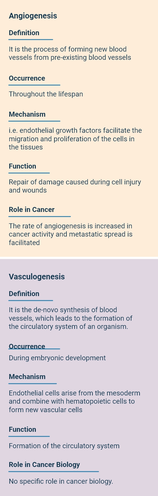
Angiogenesis, or the formation of new blood vessels, involves the strict regulation of multiple signaling pathways through which newly formed blood vessels emerge from the endothelial cells of pre-existing ones such as arteries, veins, and capillaries. Angiogenesis primarily occurs during embryogenesis and vessel reproduction in the form of vasculogenesis. At the same time, it can also be viewed as a salient process in different pathologic conditions, including cancer and inflammation, throughout the lifespan of an organism.
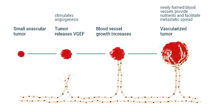
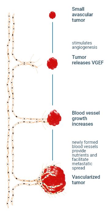
Ongoing angiogenesis is even considered an indication of cancer. In fact, the vascular endothelial growth factor (VEGF) pathway plays a pivotal role in tumor angiogenesis. Many cancers exploit this angiogenic activity to stimulate tumor growth and supply nutrients to the tumor.
Furthermore, tumor angiogenesis can result in cancer cell invasion and metastasis and plays an important role in the regulation of cancer progression, although the exact mechanism is not completely understood yet (Lugano et al., 2020).
The study of angiogenesis is a crucial part of tumor research because it can help reduce both morbidity and mortality from carcinomas. The discovery of angiogenic inhibitors in particular can help prevent neogenic blood vessel formation and tumor cell proliferation (Jiang et al., 2020). There are several successful inhibitory compounds that have been developed for various medical purposes. One notable example is Bevacizumab, which is an anti-angiogenic drug used in the treatment of various cancers, including colorectal-, lung-, and kidney cancer, among others. Bevacizumab has an inhibitory effect on the activity of vascular endothelial growth factor (VEGF), thereby reducing the formation of new blood vessels in tumors (Haibe et al., 2020). There are also several ongoing clinical trials investigating and evaluating anti-angiogenic agents in combination with immune checkpoint inhibitors (ICIs) in solid tumors (Lopes-Coelho et al., 2021).
The role of vascular endothelial growth factors (VEGF) in angiogenic processes
The major physical causes of angiogenic processes in fully developed organisms are conditions like tissue ischemia, hypoxia, inflammation, and stress. There are a number of specific factors released by tumor cells known to stimulate or inhibit angiogenesis over time, including vascular growth factors, tumor angiogenesis growth factors, inflammatory cytokines, etc.
VEGF – vascular endothelial growth factors – and VEGF receptors are a part of the major angiogenesis signaling pathways. There are five VEGF glycoproteins, which can be distinguished, namely VEGF-A, VEGF-B, VEGF-C, VEGF-D, and VEGF-E (Lee et al., 2015).
The placental growth factors PLGF 1 and 2 are also a part of the VEGF family. VEGF-A and its receptors KLT/VEGFR1 and VEGFR-2 (a tumor angiogenesis receptor) are considered to be the main target areas of antiangiogenic compounds. VEGF-A, for example, can be targeted by applying a specific therapeutic agent to inhibit microvessel growth.
Some articles suggest that VEGF may also have an additional effect on cancer progression due to the autocrine stimulation of VEGF receptors in tumor cells. There is increasing evidence of the presence of VEGFRs in liquid and solid tumor cells, e.g. in melanoma, prostate cancer, breast cancer, as well as in leukemia (Lee et al., 2015).
However, the relevance of this expression pattern is still subject to further studies. Tumor growth might not only occur due to angiogenesis induced by VEGF but can also be the result of direct stimulation via VEGFRs. Thus, endothelial cell-independent pathways may serve as the basis for useful future targets of cancer therapy methods that reach far beyond vascular endothelial growth factors (Lee et al., 2015).
Browse our IKOSA Prisma portfolio to find the right software application for your research.
The role of angiogenic markers in angiogenesis quantification
Several endothelial cell markers (e.g. PECAM-1/CD31, CD34, vWF) and angiogenesis protein markers are commonly used in immunohistochemistry (IHC) stains of human FFPE tumor sections. Quantitative data obtained from angiogenesis models can include the endothelial cell count or the expression levels of proteins associated with neovascularization.
Angiogenesis markers are measured with standardized angiogenic protein assays on the basis of particular clinical parameters like VEGF levels (Rykala et al., 2011). Specialized image analysis software also plays a central role in the quantification of angiogenic protein markers. Such tools are commonly used for molecular tumor profiling, monitoring tumor progression, and estimating tumor malignancy. In addition to its clinical uses, automated IHC quantification software has proven to be an invaluable tool in a variety of experimental models for the study of pathological angiogenesis (Kuri et al., 2022).
Angiogenesis inhibition in cancer treatment
Angiogenesis inhibitory factors serve as cancer-fighting agents by interfering with various steps in blood vessel growth. For example, they block the formation and growth of new vasculature that supports tumor progression.
Angiogenesis inhibitors may be used as monotherapy or in combination with other anti-cancer drugs. However, preclinical and clinical studies have shown higher therapeutic efficiency using a combined treatment regime in contrast to individual treatments (El-Kenawi & El-Remessy, 2013).
How do Angiogenesis Inhibitors Work
Numerous angiogenesis inhibitors are presently utilized in the management of various cancer types (Petrovic et al., 2016; Goel and Mercurio, 2014). Angiogenesis inhibitors can be classified into direct inhibitors, which target endothelial cells in the growing vasculature, or indirect inhibitors, which block the activity and expression of angiogenesis inducers. Indirect inhibitors include therapeutic compounds against oncogenes, conventional chemotherapeutic agents, or other drugs targeting various cells of the tumor microenvironment (El-Kanawi & El-Remessy, 2013).
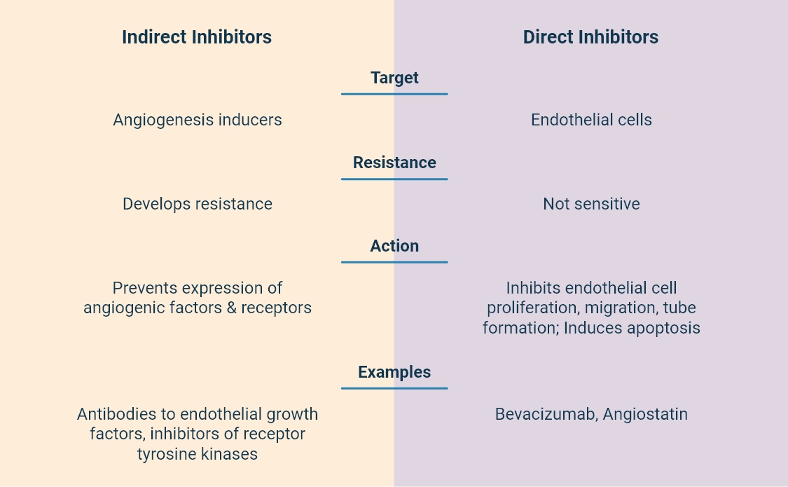
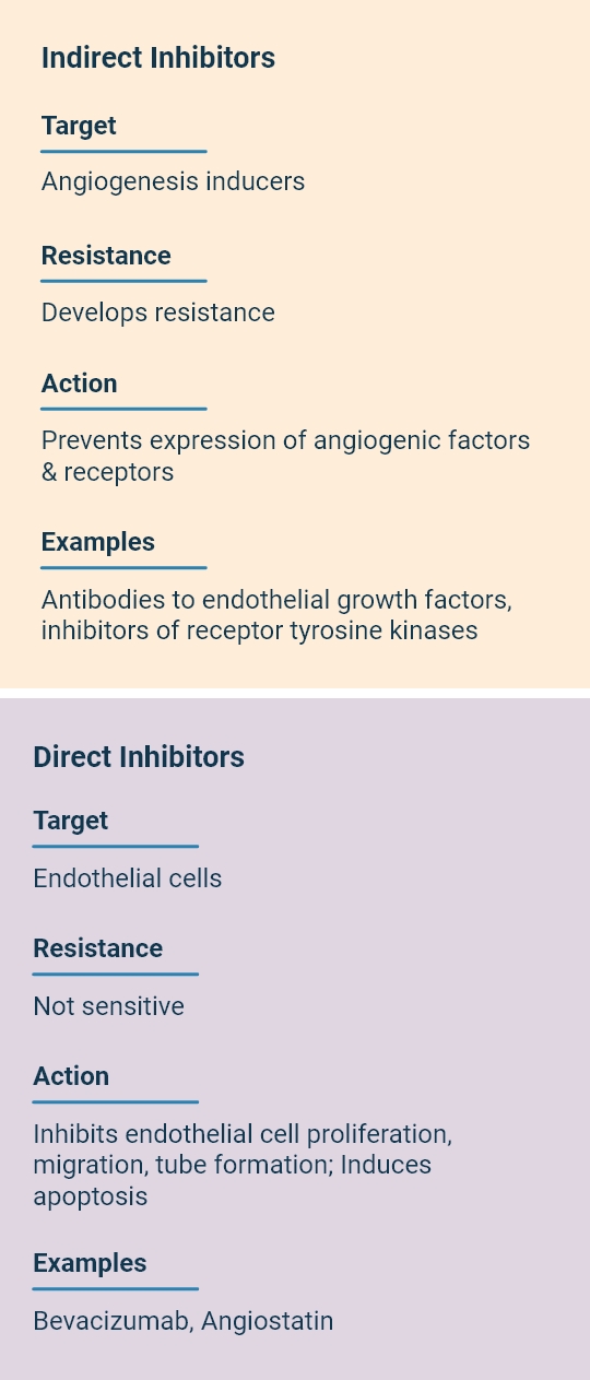
The suppression of vascular endothelial growth factors (VEGF) is described in a large body of scientific articles. This approach includes not only direct anti-VEGF treatments, either alone or in combination with chemotherapy but also immunomodulatory drugs and receptor tyrosine kinase inhibitors, targeting VEGF receptors and their signaling pathways.
Compounds that play a role in angiogenic inhibition
Among the most commonly used VEGF-targeting inhibitory agents are Avastin (Bevacizumab), Aflibercept (Zaltrap), and Ramucirumab (Cyramza) (Ramjiawan et al., 2017). Existing research gives insights into the antiangiogenic effect of novel angiogenic inhibitors. For example, promising preclinical studies revealed that Cilengitide, a selective integrin inhibitor, reduces vascular density, and vascular permeability while increasing survival rates in orthotopically-implanted glioblastoma rat models. Inhibition of FGFR-1–4, PDGFRβ, and VEGFR-1–3 with Dovitinib demonstrated anti-tumor activity in xenograft models of renal cell carcinoma (Ramjiawan et al., 2017). Current clinical studies target multiple elements within angiogenic pathways, which could potentially offer a solution for anti-angiogenic treatments. For example, Lenvatinib, a promising multi-kinase inhibitor, has demonstrated effectiveness in treating renal cell carcinoma, differentiated thyroid cancer, and hepatocellular carcinoma, primarily due to its anti-angiogenic properties. It targets key receptors including VEGFR, FGFR, PDGFRα, KIT, and RET. Lenvatinib has come a long way from research to clinical uses (Capozzi et al., 2019).
How angiogenesis research benefits from automated image analysis software
Using an elaborate angiogenesis analysis model allows researchers to examine the effects of stimulatory and inhibitory agents on vascular formation and growth.
In vitro angiogenic assays are performed on cell culture models and used to examine specific functions and processes. These assays can be classified into categories such as:
- endothelial proliferation models,
- endothelial migration models and
- endothelial cell differentiation models.
Explore our automated image analysis solutions.
In vivo assays provide a more thorough assessment of essential angiogenesis quantification parameters than in vitro and ex vivo assays, since they allow researchers to study angiogenesis dynamics in a living organism.
Ex vivo assays make use of organ or embryo culture to examine elaborate angiogenic processes. These models are more complex than in vitro assays since they closely resemble the actual biological environment and involve the interaction of vascular structures with different organ cells and surrounding tissue besides endothelial cells.
Table 1 displays an overview of the most common types of in vitro, in vivo, and ex vivo angiogenic assays discussed in recent research literature.
| In vitro assays | In vivo assays | Ex vivo assays |
|---|---|---|
| Boyden Chamber Assay | Matrigel Plug Assay | Rat Aortic Ring Assay |
| Endothelial Tube Formation Assay (EFTA) | Corneal Micropocket assay | Chick Aortic Arch Assay |
| Phagokinetic Track Assay | Chick Chorioallantoic Membrane (CAM) Assay | Choroid Sprouting Assay |
| MTT Assay | Hindlimb Ischemia Assay | Retina Model Assay |
| Matrix Invasion Assay | Zebrafish Assay | Human Placental Vessels Assay |
| Fibrin Bead Assay | Disc Assay (DAS) | Skeletal Muscle Explant Assay |
| Matrix Metalloproteinase (MMP) Assay | Sponge Implantation Method | Bovine/Murine Retinal Explant Assay |
How to choose the angiogenesis assay that fits your needs?
There is not a single universal angiogenesis assay applicable to every research design as the specifics of each method require a different procedure. Due to the heterogeneity and diversity of tissues and the complexity of angiogenic response, it seems to be an uphill task to develop a single assay suitable for all experimental designs (Shahid et al., 2017).
The factors to be considered in your research will vary depending on the purpose and the specific aspect of angiogenesis you wish to investigate, as well as on the types of cells that need to be included.
Using a variety of angiogenesis assays we can assess whether certain substances promote or inhibit blood vessel development by looking at their impact on the growth, movement, and tube formation of endothelial cells (Stryker et al., 2019). For this reason, in vitro and in vivo models are used. In vitro assays allow early-stage evaluations based on spheroid cell culture models, while in vivo methods offer a living microenvironment for exploring angiogenic mechanisms. Here are some tips on choosing the right assay based on the current state of research.
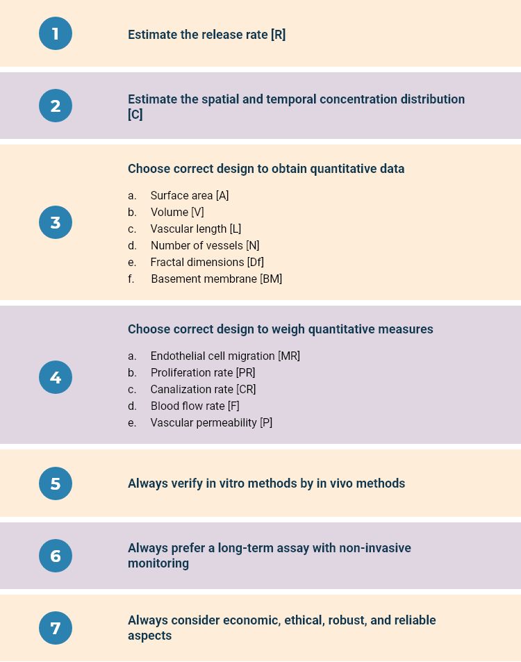
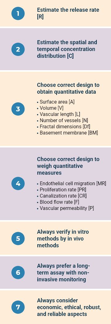
First of all, in order to evaluate dose-response curves the release rate [R] and the spatial and temporal concentration distribution [C] of the added compounds need to be estimated with the help of the chosen assay. The method you choose has to yield information on oncogene expression and angiogenic growth factor levels.
Next, the assays must be designed in a way that the quantitative parameters of the newly formed vessels can be estimated. This means the chosen methodology must enable you to obtain quantitative data on aspects of vascular morphology such as surface area [A], volume [V], vascular length [L], number of vessels in the network [N], fractal dimensions of the network [Df], and extent of basement membrane [BM].
In addition, the design of the assay should allow for weighing quantitative measures of characteristics of new vessels such as endothelial cell migration [MR], proliferation rate [PR], canalization rate [CR], blood flow rate [F], and vascular permeability [P]. It is also vital that a clear boundary between a newly formed vessel and the parent vessels can be detected with the help of the assay.
When doing the assessment, in vitro models must always be verified by subsequent in vivo studies and an angiogenesis assay for long-term and non-invasive monitoring should preferably be chosen. When opting for a particular assay, economic-, ethical-, robustness-, and reliability aspects need to be considered as well. (Shahid et al., 2017; Norrby, 2006).
Tips and tricks
With the help of advanced deep learning solutions, you can fully automate complete angiogenic analysis workflows and obtain quantitative data on vascular formation processes and markers.
Such software products largely increase throughput and reproducibility in angiogenesis image analysis. Below we look at five IKOSA image analysis solutions, which will help you obtain the optimal results while conducting the angiogenic assay of your choice. Each of these image analysis applications relies on a powerful deep learning model that is able to quickly and reliably process huge amounts of microscopy image data.
Unveiling the IKOSA portfolio of specialized solutions for the analysis of angiogenesis images
The IKOSA Platform offers a high-performance framework that will help you streamline different aspects of angiogenesis quantification. Each of the specialized software products we present to you provides added value in the evaluation of angiogenesis image data from a specific assay method.
Tap the potential of IKOSA in the analysis of CAM Assay Images
The Chorioallantoic Membrane (CAM) method is widely used in ex ovo research to quantify neovascularization and to study vascular growth patterns in the membrane lining developed around a chicken embryo on the inner surface of an eggshell. Moreover, the CAM Assay is utilized in-vivo cancer- and wound-healing research for the quantitative analysis of the angiogenic and anti-angiogenic processes.
The IKOSA CAM Assay App enables researchers to automatically extract information on morphological and spatial parameters of the vascular area on the chorioallantoic membrane, such as:
- vessel total area,
- vessel total length,
- vessel mean thickness,
- and number of branching points.
The IKOSA CAM Assay application is future-proof in this regard as it allows flexibility.
Delve into the CAM Assay and its reviews by respected institutions. Begin your exploration today!
We developed this state-of-the-art software tool for the analysis of CAM images in close cooperation with leading scientists from the Institute for Molecular and Cellular Anatomy at the University of Regensburg, the Otto Loewi Research Center at the Medical University of Graz, and the Ludwig Boltzmann Institute for Experimental and Clinical Traumatology in Vienna.
We’d like to express a special thanks to researchers Dr. Silke Härteis and Dr. Nassim Ghaffari Tabrizi-Wizsy for their valuable input.
Learn how using the IKOSA CAM Application was put to practice in an article on the quantification of tumor-induced angiogenesis in a 3D in vivo tutor model. One further publication of the research group showcases the use of our CAM Assay Application for quantifying renal cyst growth in kidney tissue.
Our cooperation with the team of Dr. Nassim Ghaffari Tabrizi-Wizsy at the Medical University of Graz gave rise to another automated tool for quantifying vasculature development in CAM images. The IKOSA CAM Grid Assay Application has been especially created for the segmentation of new blood vessels on a chorioallantoic membrane placed on a polymerized grid. Using this method allows you to collect quantitative data on parameters such as:
- number of vessels,
- total vessel area,
- mean vessel area,
- median vessel area,
- and mean image intensity.
Explore blood vessel segmentation in ex-vivo angiogenesis research with ‘onplants’ on CAM assay.
Optimize the analysis of angiogenic sprouting
Angiogenic sprouting refers to the morphogenesis of hierarchical networks of vascular sprouts such as arterioles, venules, and highly branched capillaries providing efficient blood flow to body organs. Angiogenic sprouting models are widely applied by researchers to examine the dynamics of cancer cell invasion during blood vessel sprouting in vitro studies. The spheroid sprouting assay makes use of endothelial cell spheroid culture or tumor organoids to study this process.
These methods are used to quantify the migration of cells as an indicator of angiogenic response. For this purpose, spheroids are embedded in a collagen, matrigel, or fibrin medium matrix. The migration of cells into the medium involves either the formation of single-cell sprouts or of complex capillary-like structures.
The IKOSA Spheroid Sprouting Assay Application enables the investigation of critical sprouting parameters of endothelial cell spheroids using time-lapse images. This allows you to extract spatial and temporal information on angiogenesis sprouting mechanisms. The application is perfectly suited for the quantification of features such as:
- number of sprouts,
- sprouts’ total length,
- sprouts’ total area,
- body area,
- and body circularity.
This unique spheroid sprouts analysis solution is the result of our collaboration with the Department of Obstetrics and Gynecology at the Medical University of Graz and the Ocular Angiogenesis Group at the Department of Medical Biology of Amsterdam UMC. The development of this application wouldn’t have been possible without the assistance of researchers Dr. Ursula Hiden, Jasmin Strutz MSc, and Dr. Ingeborg Klaassen.
Our cooperation partners have a proven record of scientific articles on endothelial cell response during angiogenesis including studies on:
- VEGFA signaling in human endothelial tip cells and non-tip cells
- The effect of apelin signaling in sprout progression
- Novel tip cell genes in microvascular endothelial cell monolayers
- The anti-angiogenic effect of crenolanib on cell viability, migration, sprouting, apoptosis and mitosis
- The regulatory effect of IGF-binding proteins 3 and 4 on angiogenic sprouting
Easily detect and quantify sprouts with our Spheroid Sprouting Assay.
Illuminate the mechanisms of fibrin tube formation with AI
Endothelial cell culture techniques are extensively employed for investigating the progression of vessel-like structures or tubes as part of vascular network formation studies over time.
The Endothelial Tube Formation Assay (ETFA) is a widely accepted approach for assessing the capillary-like expansion of endothelial cells on a fibrin matrix.
Therefore, EFTA is a popular in vitro method, applied in experimental wound healing and angiogenesis research to study the induction or inhibition of tube formation.
Matrigel is a solid basement membrane matrix typically utilized in the Tube Formation Assay, helping endothelial cells in the formation of tube-like structures and differentiation. This method is typically used to study the effects of various compounds on tube formation and for morphological characterization.
AI-driven image analysis software allows researchers to automatically detect and quantify extremities, branch structures, segments, and junctions of an endothelial cell tubular network.
Specifically designed for that purpose, the IKOSA Fibrin Tube Formation Assay application will help researchers gain valuable insights into central parameters such as:
- number of tubes,
- total tubes area,
- total tubes length,
- number of tube branching points,
- number of loops,
- and total loop area.
We’d like to thank Dr. Ursula Hiden and Jasmin Strutz MSc from the Department of Obstetrics and Gynecology at the Medical University of Graz for their support on this project. Find out how their team applied the Fibrin Tube Formation Assay in an article examining outgrowth, proliferation, viability, angiogenesis, and phenotype of primary human endothelial cells.
Detect and quantify endothelial cell tubes with our Fibrin Tube Formation Assay.
Streamline the analysis of vascular network formation
Angiogenesis network formation research can greatly benefit from automated deep learning applications. For instance, these applications support scientific studies on how to block new blood vessel formation in order to suppress tumor growth. In other words, researchers are seeking strategies to cut the adequate nutrient supply in the vascular network of cultured endothelial tissue.
Developed in collaboration with the Department of Obstetrics and Gynecology at the Medical University of Graz, the IKOSA Network Formation Assay application allows users to automatically collect relevant information on multiple quantitative parameters such as:
- number of tubes,
- number of branching points,
- total areas covered by cells or tubes,
- and total tube length.
Find out how the research team at the Medical University of Graz applies the Network Formation Assay for studying placental angiogenesis.
Detect branching points, loops, and cell coverage effortlessly with our Network Formation Assay App.
Did you know?
The IKOSA Platform is always evolving, so keep checking our Prisma portfolio for new angiogenesis quantification apps. If you still haven’t found a suitable software tool for the purposes of your study, consider using IKOSA AI. With the help of IKOSA AI, you can create your own image analysis application for the automated analysis of various microscopy images. Angiogenesis images make no exception, so give it a try!
Acknowledgments
We would like to thank the following project team for the opportunity to use CAM Assay images in this article:
Dr. Nassim Ghaffari Tabrizi-Wizsy (Otto Loewi Research Center, Immunology and Pathophysiology, Medical University of Graz) provided the expertise of working with the CAM Assay. Lorenz Faihs performed the experiments and imaging/analysis, as well as DI Dr. Peter Dungel and A.o. Univ.-Prof. Mag. DDr. Kurt Schicho (Ludwig Boltzmann Institute for Experimental and Clinical Traumatology and University Clinic for Cranio-, Maxillofacial and Oral Surgery, Medical University of Vienna), planned the project.
Our authors:

Lead content writer, life science professional, and simply a passionate person about technology in healthcare.

Sales manager, biomedical engineer, specialized in tissue engineering, and life science expert.

Content writer focused on the technological advancements in healthcare such as digital health literacy and telemedicine.

Health communications and marketing expert dedicated to delivering the latest topics in life science technology to healthcare professionals.
References
Berndsen, R. H., Castrogiovanni, C., Weiss, A., Rausch, M., Dallinga, M. G., Miljkovic-Licina, M., … & Nowak-Sliwinska, P. (2019). Anti-angiogenic effects of crenolanib are mediated by mitotic modulation independently of PDGFR expression. British journal of cancer, 121(2), 139-149.
Bichlmayer, E. M., Mahl, L., Hesse, L., Pion, E., Haller, V., Moehwald, A., … & Haerteis, S. (2022). A 3D In Vivo Model for Studying Human Renal Cystic Tissue and Mouse Kidney Slices. Cells, 11(15), 2269.
Capozzi, M., De Divitiis, C., Ottaiano, A., von Arx, C., Scala, S., Tatangelo, F., Delrio, P., & Tafuto, S. (2019). Lenvatinib, a molecule with versatile application: from preclinical evidence to future development in anti-cancer treatment. Cancer management and research, 11, 3847–3860. https://doi.org/10.2147/CMAR.S188316.
Dallinga, M. G., Yetkin-Arik, B., Kayser, R. P., Vogels, I., Nowak-Sliwinska, P., Griffioen, A. W., … & Schlingemann, R. O. (2018). IGF2 and IGF1R identified as novel tip cell genes in primary microvascular endothelial cell monolayers. Angiogenesis, 21(4), 823-836.
Dallinga, M. G., Habani, Y. I., Kayser, R. P., Van Noorden, C. J., Klaassen, I., & Schlingemann, R. O. (2020). IGF-binding proteins 3 and 4 are regulators of sprouting angiogenesis. Molecular Biology Reports, 47(4), 2561-2572.
Dallinga, M. G., Habani, Y. I., Schimmel, A. W., Dallinga-Thie, G. M., van Noorden, C. J., Klaassen, I., & Schlingemann, R. O. (2021). The role of heparan sulfate and neuropilin 2 in VEGFA signaling in human endothelial tip cells and non-tip cells during angiogenesis in vitro. Cells, 10(4), 926.
El-Kenawi, A.E., El-Remessy, A.B. (2013). Angiogenesis inhibitors in cancer therapy: mechanistic perspective on classification and treatment rationales. British Journal of Pharmacology, 170, 712-729.
Goel H.L., Mercurio, A.M. (2013). VEGF targets the tumour cell. Nat Rev Cancer, 13(12), 871-882.
Haibe, Y., Kreidieh, M., El Hajj, H., Khalifeh, I., Mukherji, D., Temraz, S., & Shamseddine, A. (2020). Resistance Mechanisms to Anti-angiogenic Therapies in Cancer. Frontiers in oncology, 10, 221. https://doi.org/10.3389/fonc.2020.00221.
Jiang, X., Wang, J., Deng, X., Xiong, F., Zhang, S., Gong, Z., Li, X., Cao, K., Deng, H., He, Y., Liao, Q., Xiang, B., Zhou, M., Guo, C., Zeng, Z., Li, G., Li, X., & Xiong, W. (2020). The role of microenvironment in tumor angiogenesis. Journal of experimental & clinical cancer research : CR, 39(1), 204. https://doi.org/10.1186/s13046-020-01709-5
Kuri, P. M., Pion, E., Mahl, L., Kainz, P., Schwarz, S., Brochhausen, C., … & Haerteis, S. (2022). Deep Learning-Based Image Analysis for the Quantification of Tumor-Induced Angiogenesis in the 3D In Vivo Tumor Model—Establishment and Addition to Laser Speckle Contrast Imaging (LSCI). Cells, 11(15), 2321.
Lee, S.H., Jeong, D., Han, YS., Baek, M.J. (2015). Pivotal role of vascular endothelial growth factor pathway in tumor angiogenesis. Annals of Surgical Treatment and Research, 89(1), 1-8.
Leopold, B., Strutz, J., Weiß, E., Gindlhuber, J., Birner-Gruenberger, R., Hackl, H., … & Hiden, U. (2019). Outgrowth, proliferation, viability, angiogenesis and phenotype of primary human endothelial cells in different purchasable endothelial culture media: feed wisely. Histochemistry and Cell Biology, 152(5), 377-390.
Loegl, J., Nussbaumer, E., Cvitic, S., Huppertz, B., Desoye, G., & Hiden, U. (2017). GDM alters paracrine regulation of feto-placental angiogenesis via the trophoblast. Laboratory Investigation, 97(4), 409-418.
Lopes-Coelho, F., Martins, F., Pereira, S. A., & Serpa, J. (2021). Anti-Angiogenic Therapy: Current Challenges and Future Perspectives. International journal of molecular sciences, 22(7), 3765. https://doi.org/10.3390/ijms22073765.
Lugano, R., Ramachandran, M., & Dimberg, A. (2020). Tumor angiogenesis: causes, consequences, challenges and opportunities. Cellular and molecular life sciences : CMLS, 77(9), 1745–1770. https://doi.org/10.1007/s00018-019-03351-7.
Nishida, N., Yano, H., Nishida, T., Kamura, T., Kojiro, M. (2006). Angiogenesis in cancer. Vascular Health and Risk Management, 2(3), 213-219.
Norrby, K. (2006). In vivo models of angiogenesis. J. Cell. Mol. Med., 10(3), 588-612.
Palm, M. M., Dallinga, M. G., van Dijk, E., Klaassen, I., Schlingemann, R. O., & Merks, R. M. (2016). Computational screening of tip and stalk cell behavior proposes a role for apelin signaling in sprout progression. PloS one, 11(11), e0159478.
Petrovic, N. (2016). Targeting Angiogenesis in Cancer Treatments: Where do we Stand? J Pharm Pharm Sci. 19(2), 226-238.
Ramjiawan, R.R., Griffioen, A.W., Duda, D.G. (2017). Anti-angiogenesis for cancer revisited: Is there a role for combinations with immunotherapy? Angiogenesis, 20(2), 185-204.
Rykala, J., Przybylowska, K., Majsterek, I., Pasz-Walczak, G., Sygut, A., Dziki, A., & Kruk-Jeromin, J. (2011). Angiogenesis markers quantification in breast cancer and their correlation with clinicopathological prognostic variables. Pathology & Oncology Research, 17(4), 809-817.
Shahid, I., AlMalki, W.H., AlRabia, M.W., Ahmed, M., Imam, M.T., Saifullah, M.K., Hafeez, M.H. (2017). Recent Advances in Angiogenesis Assessment Methods and their Clinical Applications, Physiologic and Pathologic Angiogenesis – Signaling Mechanisms and Targeted Therapy. Intechopen. doi:10-5772/66504.
Shih, S. C., Robinson, G. S., Perruzzi, C. A., Calvo, A., Desai, K., Green, J. E., … & Senger, D. R. (2002). Molecular profiling of angiogenesis markers. The American journal of pathology, 161(1), 35-41.
Stryker, Z. I., Rajabi, M., Davis, P.J., Mousa, S.A. (2019). Evaluation of Angiogenesis Assays. Biomedicines 7, 37, 1-13.
Zhao Y, Adjei AA. Targeting Angiogenesis in Cancer Therapy: Moving Beyond Vascular Endothelial Growth Factor. Oncologist. 2015 Jun;20(6):660-73.