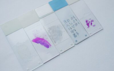
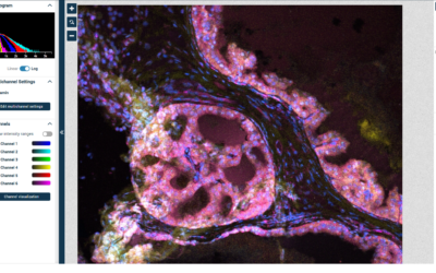
Spatial biology is a growing field in modern life science with a significant future potential. We dive deep into this exciting subject to keep you updated with the latest developments in the field. Find out about state-of-the art spatial biology...

Spatial biology is a growing field in modern life science with a significant future potential. We dive deep into this exciting subject to keep you updated with the latest developments in the field. Find out about state-of-the art spatial biology...
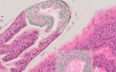
Prostate microscopy is one of the areas where the use of pathology image analysis is on the rise and about to become standard diagnostic practice. In this article, you will learn about the recent developments in automated prostate tissue analysis. Find out how...
Shifting from single marker analysis to more complex multiplex IHC analysis methods is an important step towards improving the outcomes of immunohistochemistry studies. Yet, different multiplex immunohistochemistry (mIHC) techniques generate tons of image data for...
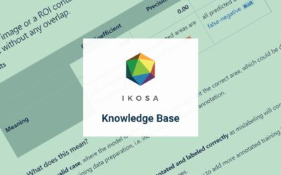
Since user education has always been a priority to us we have recently launched theIKOSA Knowledge Base - an extensive online repository of microscopy resources for an effective bioimage analysis and algorithm training with the IKOSA Platform. In...
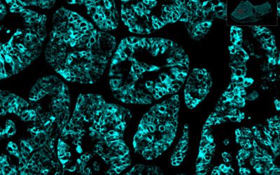
State-of-the-art nuclei segmentation techniques using deep learning technology solve many problems related to extracting reliable quantitative information on the cell nucleus level. This article presents Deep Learning (DL) based methods used for nuclei segmentation...
Learn about the latest developments in prostate tissue histology, a field in which the need for new automated image analysis methods is rapidly increasing. State-of-the-art image segmentation methods assist researchers in detecting pathological prostate conditions,...
Be the first to receive free tips about image analysis, newest articles and publications!